FREQUENTLY ASKED QUESTIONS ABOUT FETAL 2D ECHO IN PREGNANCY
ALL YOUR DOUBTS ANSWERED
What is a Fetal 2D ECHO?
Fetal 2D ECHO is an advanced ultrasound procedure designed to capture detailed images of your baby’s heart. It focuses on evaluating the heart’s chambers, valves, and blood flow, allowing doctors to detect congenital heart defects or other structural issues.
This test is often recommended when there’s a higher risk of heart abnormalities due to maternal health conditions like diabetes, family history, previous affected child or findings on routine ultrasound scanning.
Why is a Fetal 2D ECHO Performed?
A fetal 2D ECHO is performed to:
Detect congenital heart defects: Early diagnosis helps plan necessary medical interventions.
Evaluate blood flow patterns: The test examines how blood moves in and out of the baby’s heart.
Monitor fetal heart rhythm: It checks for arrhythmias or other irregularities in the heartbeat.
When is a Fetal 2D ECHO Recommended?
Doctors typically recommend a fetal 2D ECHO if:
There is a family history of congenital heart defects.
Abnormalities were detected during a routine ultrasound.
The mother has pre-existing medical conditions like diabetes, lupus, or epilepsy.
There has been an exposure to certain medications or infections during pregnancy.
The pregnancy involves twins or multiple pregnnaces.
Assisted reproductive techniques were used to conceive.
At What Stage of Pregnancy is the Test Done?
The ideal time for a fetal 2D ECHO is between 20 and 24 weeks of pregnancy. At this stage, your baby’s heart is developed enough for a thorough evaluation, but the test can be done earlier or later if necessary. The visualisation is best around 24-26 weeks of pregnancy. In earlier or later weeks, the visualisation may not be optimal. We usually club the fetal ECHO with a TIFFA Scan or Growth Scan.
How Do I Prepare for a Fetal 2D ECHO?
Preparing for this test is simple:
In most cases, no special preparation is required. It is better to have a snack or meal before the scan
You may be advised to have a moderately full bladder for better imaging in early pregnancy.
What Happens During the Procedure?
You’ll be asked to lie on an examination table.
A gel will be applied to your abdomen to help transmit the sound waves.
A transducer (a handheld device) is moved over your abdomen to capture images of your baby’s heart.
The procedure is non-invasive, painless, and usually takes 30 to 40 minutes.
If the position of the fetus is not optimal for visualisation, you may be asked to have some tea or juice so that the fetus moves and the scan will be continued.
Is Fetal 2D ECHO Safe?
Yes, fetal 2D ECHO is completely safe for both the mother and the baby. It uses high-frequency sound waves to generate images, which are non-invasive and free from radiation.
Is the Fetal Gender Revealed?
No. fetal gender determination is prohibited and is a punishable offense under the PCPNDT Act. It is not determined at this scan or for that matter during any other scan
Can Family Members View the fetal 2D ECHO Scan?
Yes, but inform the reception desk beforehand. Post-scan, family members can view the fetus and its details. However, photography and videography are strictly prohibited under the PNDT Act.
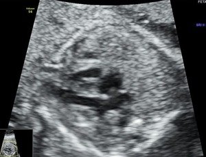
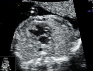
normal fetal heart
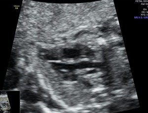
feal heart- normal LVOT
fetal heart- normal 3VT view
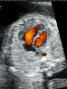
blood flow in fetal heart
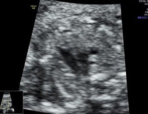
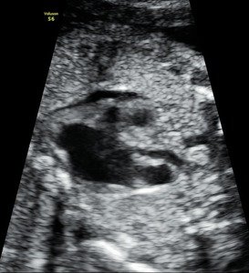
abnormal fetal heart
abnormal fetal heart
Early assessment and detection of fetal heart abnormalities can make a significant difference in ensuring your baby’s health and well-being and planning further pregnancy care and neonatal care after birth.
YOU CAN SCHEDULE YOUR FETAL 2D ECHO TODAY FOR EVALUATION OF THE FETAL HEART
We have a high level of expertise combined with a High Resolution Ultrasound Machine & a compassionate approach